Hot NewsAnatomy Of Chest : Radiology Chest Xray Normal
Anatomy Of Chest : Radiology Chest Xray Normal
Anatomy Of Chest : Radiology Chest Xray Normal. The first rib is a short, flat rib that is much wider and more curved than those previously described. (1) the pectoralis major, and (2) the pectoralis minor. Intravenous contrast is seen in the left ventricle (1) and descending aorta (2). Chest muscles anatomy (1) pectoralis major muscle. Thoracic cavity, also called chest cavity, the second largest hollow space of the body.
The circulatory system does most of its work. Chest a man's chest — like the rest of his body — is covered with skin that has two layers. The pectoralis major and the pectoralis minor, known collectively as your pecs. A line is drawn from anterior surface of the body of 6th thoracic vertebrae passing through the apex of the heart up to anterior lower most part of diaphragm. Anatomy of the thorax, heart, abdomen and pelvis recommended text gray's anatomy for students.
Thorax Chest Anatomy Archives Anatomy Note from www.anatomynote.com Applied anatomy of the chest wall and mediastinum petros mirilas michael e. Thoracic cavity, also called chest cavity, the second largest hollow space of the body. An overview of the anatomy visible in a transverse computed axial tomographical image of the thorax (and part of the abdomen) performed with intravenous cont. Browse 2,525 female chest anatomy stock photos and images available, or start a new search to explore more stock photos and images. Anatomy of the thoracic wall. Book of chest anatomy is a passive item. A line is drawn from anterior surface of the body of 6th thoracic vertebrae passing through the apex of the heart up to anterior lower most part of diaphragm. It spreads out like a fan and covers the rib cage like an armor plate.
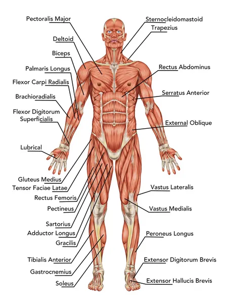
This page provides an overview of the chest muscle group.
Applied anatomy of the chest wall and mediastinum petros mirilas michael e. Related posts of anatomy of the chest area endocrine glands diagram picture. Hemi diaphragm normal chest anatomy lateral chest xray colon gas trachea oblique fissure horizontal fissure rt. The chest wall is comprised of skin, fat, muscles, and the thoracic skeleton. Strictures, acute syndromes, neoplasms and vascular impressions Browse 2,525 female chest anatomy stock photos and images available, or start a new search to explore more stock photos and images. The skeleton of the thoracic wall is formed by the twelve thoracic vertebra posteriorly, Anatomy of the thoracic wall. The major muscle in the chest is the pectoralis major. Thoracic cavity, also called chest cavity, the second largest hollow space of the body. It is enclosed by the ribs, the vertebral column, and the sternum, or breastbone, and is separated from the abdominal cavity (the body's largest hollow space) by a muscular and membranous partition, the diaphragm. Book of chest anatomy is a passive item. Chest a man's chest — like the rest of his body — is covered with skin that has two layers.
An overview of the anatomy visible in a transverse computed axial tomographical image of the thorax (and part of the abdomen) performed with intravenous cont. Here, we break down the anatomy of your chest muscles. The pectoralis major and the pectoralis minor, known collectively as your pecs. It's also sometimes referred to as the breastbone. It is enclosed by the ribs, the vertebral column, and the sternum, or breastbone, and is separated from the abdominal cavity (the body's largest hollow space) by a muscular and membranous partition, the diaphragm.
ሠChest Anatomy Stock Pictures Royalty Free Pectoralis Major Images Download On Depositphotos from st.depositphotos.com Computed tomography (ct) of the chest can detect pathology that may not show up on a conventional chest radiograph(1). Chest a man's chest — like the rest of his body — is covered with skin that has two layers. Related posts of anatomy of the chest and stomach human cellular respiration diagram. A line is drawn from anterior surface of the body of 6th thoracic vertebrae passing through the apex of the heart up to anterior lower most part of diaphragm. How to view the anatomical labels. The chest or thorax is the region between the neck and diaphragm that encloses organs, such as the heart, lungs, esophagus, trachea, and thoracic diaphragm. Endocrine glands diagram picture 13 photos of the endocrine glands diagram picture diagram showing endocrine glands, endocrine glands chart, endocrine glands diagram labeled, endocrine system diagram glands, hypothalamus diagram, pituitary gland diagram, thyroid gland diagram, human anatomy, diagram showing. Human cellular respiration diagram 8 photos of the human cellular respiration diagram aerobic cellular respiration diagram, cellular respiration diagram and explanation, cellular respiration diagram for kids, cellular respiration diagram worksheet, cellular respiration photosynthesis diagram, cellular.
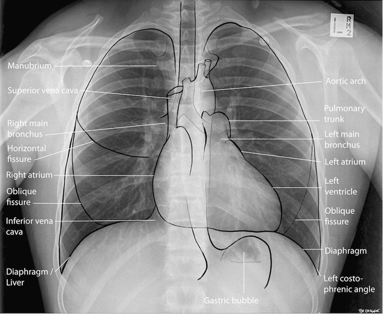
Structures of the heart such as the right ventricle (3), intraventricular septum (4), left ventricular free wall (5) and papillary muscles (arrow) are clearly seen.
An overview of the anatomy visible in a transverse computed axial tomographical image of the thorax (and part of the abdomen) performed with intravenous cont. Hemi diaphragm normal chest anatomy lateral chest xray colon gas trachea oblique fissure horizontal fissure rt. This atlas is a comprehensive and affordable learning tool for medical students and residents and especially for radiologists and pneumologists. Endocrine glands diagram picture 13 photos of the endocrine glands diagram picture diagram showing endocrine glands, endocrine glands chart, endocrine glands diagram labeled, endocrine system diagram glands, hypothalamus diagram, pituitary gland diagram, thyroid gland diagram, human anatomy, diagram showing. Human cellular respiration diagram 8 photos of the human cellular respiration diagram aerobic cellular respiration diagram, cellular respiration diagram and explanation, cellular respiration diagram for kids, cellular respiration diagram worksheet, cellular respiration photosynthesis diagram, cellular. 1 effects 2 notes 3 trivia 4 see also improves the contents of broken chests. Thoracic cavity, also called chest cavity, the second largest hollow space of the body. The chest anatomy includes the pectoralis major, pectoralis minor and the serratus anterior. (1) the pectoralis major, and (2) the pectoralis minor. Strictures, acute syndromes, neoplasms and vascular impressions Structures of the heart such as the right ventricle (3), intraventricular septum (4), left ventricular free wall (5) and papillary muscles (arrow) are clearly seen. Related posts of anatomy of the chest and stomach human cellular respiration diagram. It provides protection to vital organs (eg, heart and major vessels, lungs, liver) and provides stability for movement.
Book of chest anatomy is a passive item. An overview of the anatomy visible in a transverse computed axial tomographical image of the thorax (and part of the abdomen) performed with intravenous cont. It's also sometimes referred to as the breastbone. Structures of the heart such as the right ventricle (3), intraventricular septum (4), left ventricular free wall (5) and papillary muscles (arrow) are clearly seen. Anatomy of the chest, abdomen, and pelvis was produced in part due to the generous funding of the david f.
Anatomy Of A Chest X Ray Radiologypics Com from radiologypics.files.wordpress.com Related posts of anatomy of the chest and stomach human cellular respiration diagram. Here, we break down the anatomy of your chest muscles. The tissue adjacent to the aorta is the. The epidermis is the outermost layer that provides a protective, waterproof seal over the body. Intravenous contrast is seen in the left ventricle (1) and descending aorta (2). The circulatory system does most of its work. The dominant muscle in the upper chest is the pectoralis major. How to view the anatomical labels.
Strictures, acute syndromes, neoplasms and vascular impressions
Endocrine glands diagram picture 13 photos of the endocrine glands diagram picture diagram showing endocrine glands, endocrine glands chart, endocrine glands diagram labeled, endocrine system diagram glands, hypothalamus diagram, pituitary gland diagram, thyroid gland diagram, human anatomy, diagram showing. Swensen fund for innovation in teaching. Skandalakis chest wall embryogenesis the muscles of the chest develop from the somites found in the mesoderm. The major muscle in the chest is the pectoralis major. In particular, the right side of the chest is home to several structures including the right side of the heart, the three lobes of the right lung, the ascending aorta, the pulmonary blood vessels,. Your sternum is a bone that's located in the middle of your chest. 1 effects 2 notes 3 trivia 4 see also improves the contents of broken chests. The chest or thorax is the region between the neck and diaphragm that encloses organs, such as the heart, lungs, esophagus, trachea, and thoracic diaphragm. Studied the anatomy of the breast, its topography, innervation, vascularization and lymphatic drainage, and correlated the anatomical data with the classification of lymph node groups that is frequently utilized by mastologists. It is enclosed by the ribs, the vertebral column, and the sternum, or breastbone, and is separated from the abdominal cavity (the body's largest hollow space) by a muscular and membranous partition, the diaphragm. This page provides an overview of the chest muscle group. Related posts of anatomy of the chest area endocrine glands diagram picture. The tissue adjacent to the aorta is the.

0 comments