Hot NewsHip Muscles Diagram : Tight Hip Flexors Think Again Mana Performance Therapy Mana Performance Therapy
Hip Muscles Diagram : Tight Hip Flexors Think Again Mana Performance Therapy Mana Performance Therapy
Hip Muscles Diagram : Tight Hip Flexors Think Again Mana Performance Therapy Mana Performance Therapy. Leg hip muscles diagram / simple diagram of leg muscles.other's hip abductor muscles strengthening exercises. The muscles of the hip can be divided into three different. Related posts of muscles of the lower back and hip diagram neck muscle anatomy ultrasound. These are often divided into four groups according to their orientation around the hip joint: The view on the left has the rectus femoris cut away to show the vastus intermedius which is below it.
Large ligaments, tendons, and muscles around the hip joint hold the bones (ball and socket) in place and keep it from dislocating. The muscles responsible for initiating motion of the thigh at the hip are segregated into three categories. The iliopsoas muscle flexes your hip, bends your trunk towards your thigh and rotates your thigh bone. The hip muscles work together to carry out 4 different types of movement: Muscles play an important role in the.
4 Muscles And Insertions Of Pelvis Hip And Thigh Download Scientific Diagram from www.researchgate.net Posted on april 21, 2019april 20, 2019. These are often divided into four groups according to their orientation around the hip joint: The hip flexors can be found connecting the top of the femur, which is the largest bone in the body, to the lower back, hips, and groin. The hip joint is one of the most flexible joints in the entire human body. The hip muscles encompass many muscles of the hip and thigh whose main function is to act on the thigh at the hip joint and stabilize the pelvis.without them, walking would be impossible. Rectus femoris muscle, one of the quadriceps muscles on the front of your thigh. The four groups are the anterior group, the posterior group, adductor group. This diagram with labels depicts and explains the details of hip muscles diagram.
In human anatomy, the muscles of the hip joint are those muscles that cause movement in the hip.
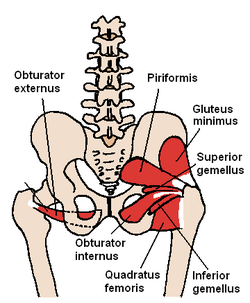
Muscles of the hip & thigh (quadriceps, hips). When hip tendonitis is a pain. Inner hip muscles there are nine inner hip muscles, found at the anterior side of the pelvis: These muscles can be grouped based upon their location and function. The quadriceps group of four muscles. Bones, joints, muscles | kenhub. Iliacus, psoas major, psoas minor, obturator externus, obturator internus, superior and inferior gemelli, piriformis, and quadratus femoris muscles. This diagram with labels depicts and explains the details of hip muscles diagram. The superficial muscles of the thigh. The posterior and lateral muscles the posterior and lateral muscles are also referred to as the muscles of the buttocks. The muscles which make up this group include the gluteus minimus, the gluteus maximus, the gluteus medius, and the tensor fasciae latae. Iliopsoas muscle, a hip flexor muscle that attaches to the upper thigh bone. Hip pain on the outside of your hip, upper thigh or outer buttock is usually caused by problems with muscles, ligaments, tendons and other soft tissues that surround your hip joint.
These muscles can be grouped based upon their location and function. Smartdraw includes 1000s of professional healthcare and anatomy chart templates that you can modify and make your own. The muscles in the hip are responsible for the movement of the hip and, by proxy, the leg. You can strain or tear your hip flexor muscles through sudden movements or falls. Leg hip muscles diagram / simple diagram of leg muscles.other's hip abductor muscles strengthening exercises.
External Obturator Muscle Wikipedia from upload.wikimedia.org When hip tendonitis is a pain. Iliacus, psoas major, psoas minor, obturator externus, obturator internus, superior and inferior gemelli, piriformis, and quadratus femoris muscles. Snapping hip syndrome is the result of the iliopsoas tendon subluxing over the greater trochanter or ilopectineal eminence. Ligaments are soft tissue structures that connect bones to bones.a joint capsule is a watertight sac that surrounds a joint.in the hip, the joint capsule is formed by a group of three strong ligaments that connect the femoral head to the acetabulum. You can strain or tear your hip flexor muscles through sudden movements or falls. The hip flexors can be found connecting the top of the femur, which is the largest bone in the body, to the lower back, hips, and groin. Repeated strains in muscles about the hip and pelvis may be associated with athletic pubalgia (also called sports hernia). The many muscles of the hip provide movement, strength, and stability to the hip joint and the bones of the hip and thigh.
Iliacus, psoas major, psoas minor, obturator externus, obturator internus, superior and inferior gemelli, piriformis, and quadratus femoris muscles.
Smartdraw includes 1000s of professional healthcare and anatomy chart templates that you can modify and make your own. In human anatomy, the muscles of the hip joint are those muscles that cause movement in the hip. Large ligaments, tendons, and muscles around the hip joint hold the bones (ball and socket) in place and keep it from dislocating. When hip tendonitis is a pain. This condition is discussed in sports hernia (athletic pubalgia). Muscles of the hip & thigh (quadriceps, hips). The muscles work together to enable movement and keep the hip in alignment. Iliopsoas muscle, a hip flexor muscle that attaches to the upper thigh bone. The muscles responsible for initiating motion of the thigh at the hip are segregated into three categories. The hip muscles encompass many muscles of the hip and thigh whose main function is to act on the thigh at the hip joint and stabilize the pelvis.without them, walking would be impossible. The iliopsoas muscle flexes your hip, bends your trunk towards your thigh and rotates your thigh bone. These are often divided into four groups according to their orientation around the hip joint: A sports hernia is a strain or tear of any soft tissue (muscle, tendon, ligament) in the lower abdomen or groin area.
Lunges that relieve back pain low back pain program. Human muscles enable movement it is important to understand what they do in order to diagnose sports the movements generated at the foot and lower leg are plantar flexion (foot points down), dorsi the hip and pelvic muscles include: The piriformis is the horizontal muscle in the center of the picture running over the top of the sciatic nerve. Muscles of thigh and the hip. This is a diagram of the larger and more surface muscles of the low back.
Muscles Of Pelvis Hip And Buttock Anatomical Chart Osta International from www.osta.ca The posterior and lateral muscles the posterior and lateral muscles are also referred to as the muscles of the buttocks. This condition is discussed in sports hernia (athletic pubalgia). They allow you to move your leg or knee up towards your torso, as well as to bend your torso forward at the hip. In human anatomy, the muscles of the hip joint are those muscles that cause movement in the hip.most modern anatomists define 17 of these muscles, although some additional muscles may sometimes be considered. The many muscles of the hip provide movement, strength, and stability to the hip joint and the bones of the hip and thigh. The hip muscles work together to carry out 4 different types of movement: Muscle diagram black woman female body names muscle diagram most important muscles of an athletic black man anterior and canstock from comps.canstockphoto.com the thigh bone or femur and the pelvis join to form the hip joint. The hip joint is one of the most flexible joints in the entire human body.
This article will introduce the muscles in each group and touch on their origin, insertion, function, and innervation.
Related posts of muscles of the lower back and hip diagram muscle anatomy posterior. The quadriceps group of four muscles. Large ligaments, tendons, and muscles around the hip joint hold the bones (ball and socket) in place and keep it from dislocating. Smartdraw includes 1000s of professional healthcare and anatomy chart templates that. The hip flexors can be found connecting the top of the femur, which is the largest bone in the body, to the lower back, hips, and groin. There are various hip flexor muscles that all work to. The muscles which make up this group include the gluteus minimus, the gluteus maximus, the gluteus medius, and the tensor fasciae latae. Inner hip muscles there are nine inner hip muscles, found at the anterior side of the pelvis: 1713 x 2175 jpeg 1066 кб. Human muscle system, the muscles of the human body that work the skeletal system, that are under voluntary. The piriformis is the horizontal muscle in the center of the picture running over the top of the sciatic nerve. They can be divided into three main groups: The muscles of the hip can be divided into three different.


0 comments